Copyright
©The Author(s) 2017.
World J Gastroenterol. Dec 28, 2017; 23(48): 8452-8464
Published online Dec 28, 2017. doi: 10.3748/wjg.v23.i48.8452
Published online Dec 28, 2017. doi: 10.3748/wjg.v23.i48.8452
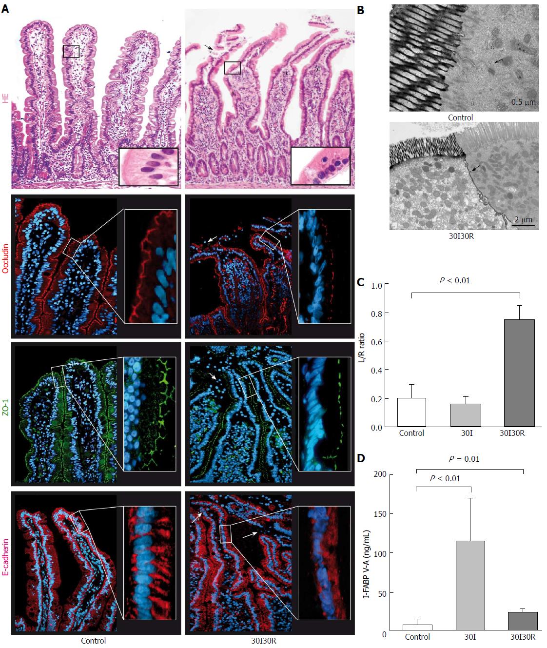
Figure 3 Thirty min of reperfusion results in tight junction damage and functional intestinal barrier loss.
A: After 30 min of reperfusion (30I30R) damaged epithelial cells were pinched off and shed into the lumen (arrow). Immunofluorescence for ZO-1 and occludin revealed irregular distribution patterns (arrowheads) and loss of expression in the epithelial sheets at the tips of the villi, together with the disruption of E-Cadherin showing only diffuse staining in the cell cytoplasm; B: Lanthanum was still able to penetrate between two adjacent enterocytes (arrowhead) demonstrating disruption of TJs and AJs at 30I30R; C: Plasma L/R ratio was significantly elevated at 30I30R compared to control (0.75 ± 0.10 vs 0.20 ± 0.09, P < 0.01), indicating increased intestinal permeability after short reperfusion; D: Plasma I-FABP gradually reduce following 30I30R but was still significantly elevated compared to control (27.06 ± 2.73 ng/mL vs 1.75 ± 0.78 ng/mL, P = 0.01), indicating entrocyte membrane integrity loss. Electron microscopy scale bars = 0.5 μm and 2 μm respectively.
- Citation: Schellekens DH, Hundscheid IH, Leenarts CA, Grootjans J, Lenaerts K, Buurman WA, Dejong CH, Derikx JP. Human small intestine is capable of restoring barrier function after short ischemic periods. World J Gastroenterol 2017; 23(48): 8452-8464
- URL: https://www.wjgnet.com/1007-9327/full/v23/i48/8452.htm
- DOI: https://dx.doi.org/10.3748/wjg.v23.i48.8452


