Copyright
©The Author(s) 2017.
World J Gastroenterol. Dec 28, 2017; 23(48): 8452-8464
Published online Dec 28, 2017. doi: 10.3748/wjg.v23.i48.8452
Published online Dec 28, 2017. doi: 10.3748/wjg.v23.i48.8452
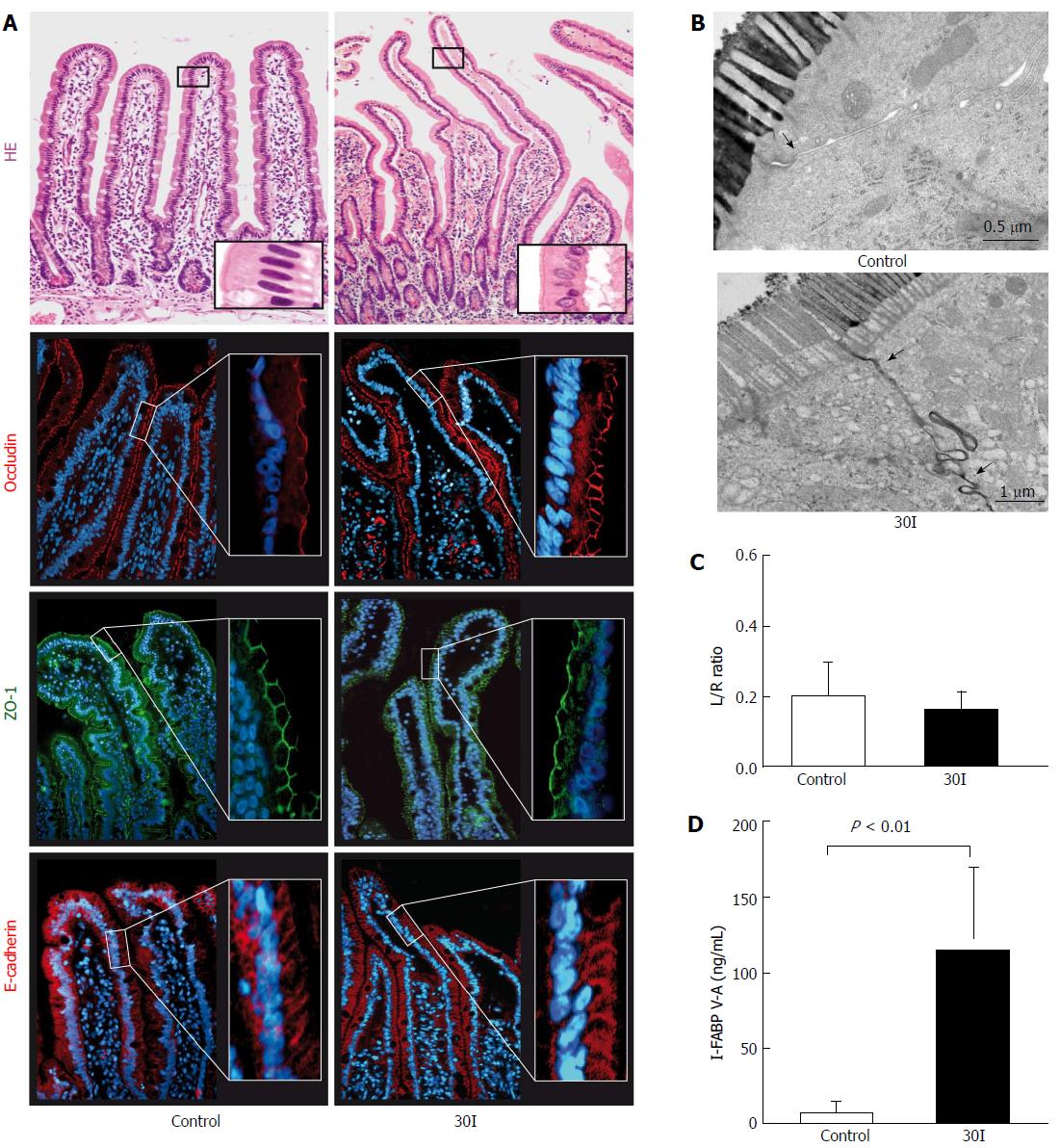
Figure 1 Changes in intestinal barrier function after 30 min of ischemia.
A: HE staining showed an intact epithelial lining in control tissue. After 30 min of ischemia (30I) subepithelial spaces were visualized at the tips of the villi. Strong and evenly distributed immunofluorescent stainings for ZO-1, occludin and E-Cadherin were observed, over the tips of the villi, in both control tissue and jejunum subjected to 30I; B: Electron microscopic images of two enterocytes with interconnecting tight junctions demonstrate in control tissue that tight junctions are intact (arrow) and the contrast dye lanthanum remains intraluminally. On the contrary, after 30I, lanthanum is able to penetrate the paracellular space, indicating tight junction integrity loss (arrow); C: Plasma L/R ratio’s however, remained unchanged after ischemia when compared with control; D: Mean plasma I-FABP V-A concentration differences show a significant release after ischemia when compared with control (126.6 ng/mL ± 65.53 ng/mL vs 1.75 ng/mL ± 0.78 ng/mL, P < 0.01), reflecting enterocyte damage. Electron microscopy scale bars = 0.5 μm and 1 μm respectively.
- Citation: Schellekens DH, Hundscheid IH, Leenarts CA, Grootjans J, Lenaerts K, Buurman WA, Dejong CH, Derikx JP. Human small intestine is capable of restoring barrier function after short ischemic periods. World J Gastroenterol 2017; 23(48): 8452-8464
- URL: https://www.wjgnet.com/1007-9327/full/v23/i48/8452.htm
- DOI: https://dx.doi.org/10.3748/wjg.v23.i48.8452


