Copyright
©The Author(s) 2017.
World J Gastroenterol. Dec 21, 2017; 23(47): 8367-8375
Published online Dec 21, 2017. doi: 10.3748/wjg.v23.i47.8367
Published online Dec 21, 2017. doi: 10.3748/wjg.v23.i47.8367
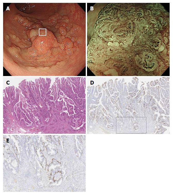
Figure 6 Endoscopic and histologic features of a lesion with irregular white opaque substance.
A: Colonoscopy shows a protruding lesion in the rectum; B: Magnifying endoscopic findings with narrow-band imaging of box in Figure 6a show irregular WOS. WOS is disorganized and asymmetrical; C: Histological examination of the resected specimen shows well to moderately differentiated adenocarcinoma invading the deep submucosal layer (invasion depth; 4,320 μm); D: A low-power view with adipophilin staining shows that the depth of adipophilin expression is deep; E: A high-power view of the box in Figure 6d shows that adipophilin is detected within the neoplastic epithelium. WOS: White opaque substance.
- Citation: Kawasaki K, Eizuka M, Nakamura S, Endo M, Yanai S, Akasaka R, Toya Y, Fujita Y, Uesugi N, Ishida K, Sugai T, Matsumoto T. Association between white opaque substance under magnifying colonoscopy and lipid droplets in colorectal epithelial neoplasms. World J Gastroenterol 2017; 23(47): 8367-8375
- URL: https://www.wjgnet.com/1007-9327/full/v23/i47/8367.htm
- DOI: https://dx.doi.org/10.3748/wjg.v23.i47.8367


