Copyright
©The Author(s) 2017.
World J Gastroenterol. Dec 21, 2017; 23(47): 8367-8375
Published online Dec 21, 2017. doi: 10.3748/wjg.v23.i47.8367
Published online Dec 21, 2017. doi: 10.3748/wjg.v23.i47.8367
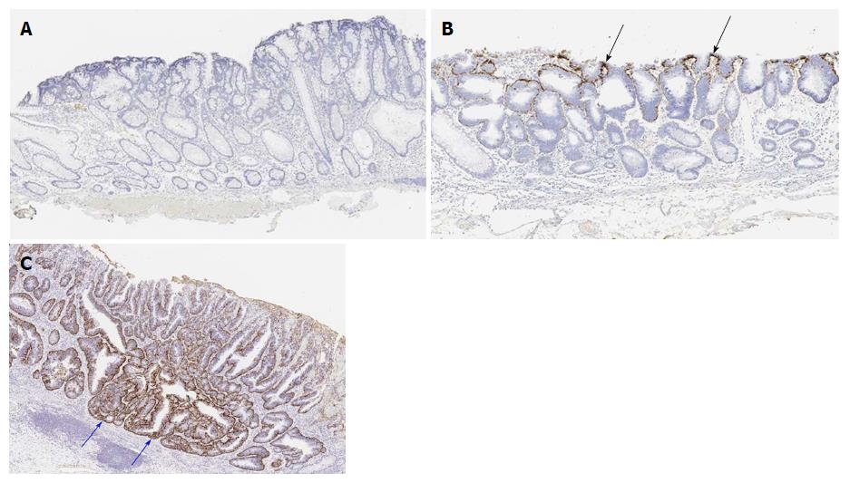
Figure 2 Histopathological findings: Adipophilin immunostaining.
A: Score 0, adipophilin is not detected within the neoplastic epithelium; B: Score 1, adipophilin is detected within the neoplastic epithelium. The depth of adipophilin expression is superficial (black arrows); C: Score 2, adipophilin is detected within the neoplastic epithelium. Adipophilin expression is deep (blue arrows).
- Citation: Kawasaki K, Eizuka M, Nakamura S, Endo M, Yanai S, Akasaka R, Toya Y, Fujita Y, Uesugi N, Ishida K, Sugai T, Matsumoto T. Association between white opaque substance under magnifying colonoscopy and lipid droplets in colorectal epithelial neoplasms. World J Gastroenterol 2017; 23(47): 8367-8375
- URL: https://www.wjgnet.com/1007-9327/full/v23/i47/8367.htm
- DOI: https://dx.doi.org/10.3748/wjg.v23.i47.8367


