Copyright
©The Author(s) 2017.
World J Gastroenterol. Dec 14, 2017; 23(46): 8256-8260
Published online Dec 14, 2017. doi: 10.3748/wjg.v23.i46.8256
Published online Dec 14, 2017. doi: 10.3748/wjg.v23.i46.8256
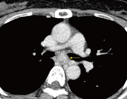
Figure 2 Computed tomography images.
Computed tomography reveals a well demarcated, heterogeneous, esophageal tumor in the mid-thoracic esophagus (arrow). The longitudinal diameter of the tumor was 60 mm.
- Citation: Onodera Y, Nakano T, Takeyama D, Maruyama S, Taniyama Y, Sakurai T, Heishi T, Sato C, Kumagai T, Kamei T. Combined thoracoscopic and endoscopic surgery for a large esophageal schwannoma. World J Gastroenterol 2017; 23(46): 8256-8260
- URL: https://www.wjgnet.com/1007-9327/full/v23/i46/8256.htm
- DOI: https://dx.doi.org/10.3748/wjg.v23.i46.8256


