Copyright
©The Author(s) 2017.
World J Gastroenterol. Dec 7, 2017; 23(45): 8097-8103
Published online Dec 7, 2017. doi: 10.3748/wjg.v23.i45.8097
Published online Dec 7, 2017. doi: 10.3748/wjg.v23.i45.8097
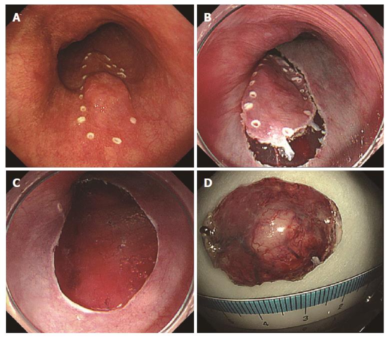
Figure 2 Intraoperative endoscopy.
A: Marking of lesion; B: Incision of lesion perimeter; C: Resected surface after excision; D: Excised specimen.
- Citation: Yoshikawa K, Kinoshita A, Hirose Y, Shibata K, Akasu T, Hagiwara N, Yokota T, Imai N, Iwaku A, Kobayashi G, Kobayashi H, Fushiya N, Kijima H, Koike K, Kaneyama H, Ikeda K, Saruta M. Endoscopic submucosal dissection in a patient with esophageal adenoid cystic carcinoma. World J Gastroenterol 2017; 23(45): 8097-8103
- URL: https://www.wjgnet.com/1007-9327/full/v23/i45/8097.htm
- DOI: https://dx.doi.org/10.3748/wjg.v23.i45.8097


