Copyright
©The Author(s) 2017.
World J Gastroenterol. Dec 7, 2017; 23(45): 8090-8096
Published online Dec 7, 2017. doi: 10.3748/wjg.v23.i45.8090
Published online Dec 7, 2017. doi: 10.3748/wjg.v23.i45.8090
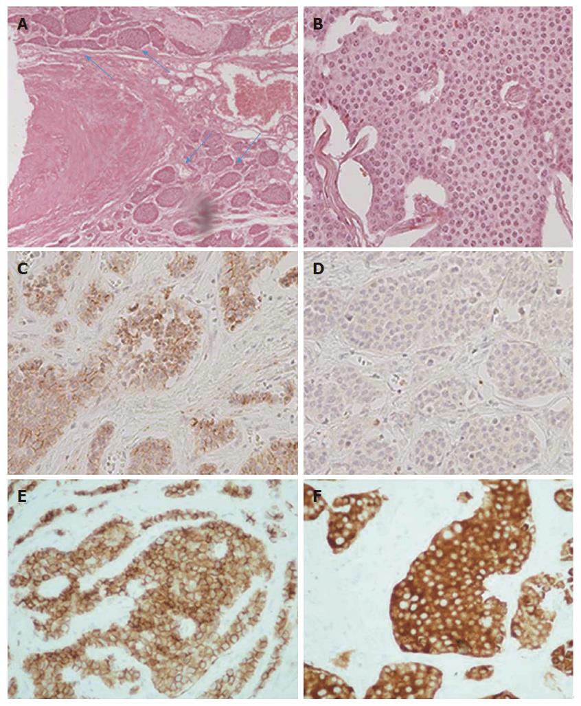
Figure 7 Histopathological findings.
A: Vascular and perineural invasion of tumor cells (blue arrows), medium magnification (100 ×); B: Tumor cells with characteristic nuclear appearance, high magnification (400 ×); C: Immunohistochemical staining reveals strong positivity for chromogranin A marker, high magnification (400 ×); D: Less than 1% of tumor cells reveal positivity for proliferative marker Ki-67, high magnification (400 ×); E: Immunohistochemical staining reveals strong positivity for CD56 marker, high magnification (400 ×); F: Immunohistochemical staining reveals strong positivity for synaptophysin marker, high magnification (400 ×).
- Citation: Mantzoros I, Savvala NA, Ioannidis O, Parpoudi S, Loutzidou L, Kyriakidou D, Cheva A, Intzos V, Tsalis K. Midgut neuroendocrine tumor presenting with acute intestinal ischemia. World J Gastroenterol 2017; 23(45): 8090-8096
- URL: https://www.wjgnet.com/1007-9327/full/v23/i45/8090.htm
- DOI: https://dx.doi.org/10.3748/wjg.v23.i45.8090


