Copyright
©The Author(s) 2017.
World J Gastroenterol. Dec 7, 2017; 23(45): 7989-7999
Published online Dec 7, 2017. doi: 10.3748/wjg.v23.i45.7989
Published online Dec 7, 2017. doi: 10.3748/wjg.v23.i45.7989
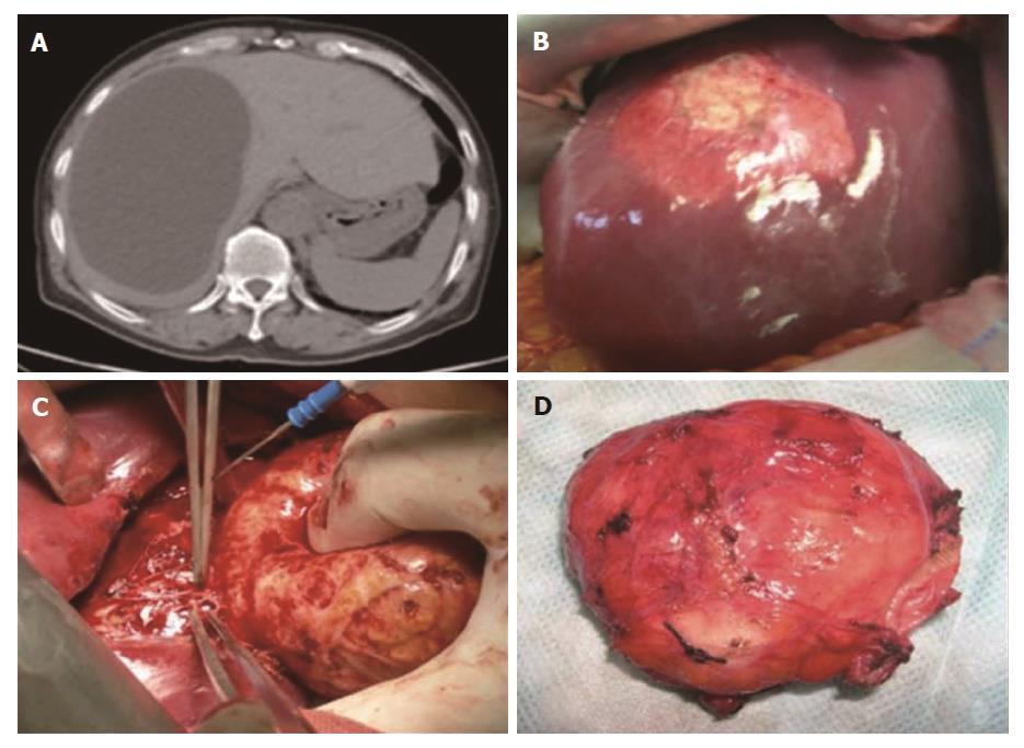
Figure 1 Echinococcus granulosus protoscolex collection.
The protoscolices were collected from patients with hepatic hydatid cysts. A: Type I hydatid cyst (WHO classification) presents as a well-defined unilocular and fluid attenuation lesion in the liver; B: The single cyst appearance during an open surgery; C: The complete removal of the hydatid cyst from the liver; D: The hydatid cyst is full of protoscolices.
- Citation: Zhang RQ, Chen XH, Wen H. Improved experimental model of hepatic cystic hydatid disease resembling natural infection route with stable growing dynamics and immune reaction. World J Gastroenterol 2017; 23(45): 7989-7999
- URL: https://www.wjgnet.com/1007-9327/full/v23/i45/7989.htm
- DOI: https://dx.doi.org/10.3748/wjg.v23.i45.7989


