Copyright
©The Author(s) 2017.
World J Gastroenterol. Dec 7, 2017; 23(45): 7978-7988
Published online Dec 7, 2017. doi: 10.3748/wjg.v23.i45.7978
Published online Dec 7, 2017. doi: 10.3748/wjg.v23.i45.7978
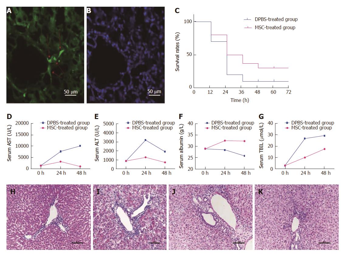
Figure 1 Survival rate and biochemical indicators in rats are improved after mesenchymal stem cell treatment.
Colonization of mesenchymal stem cells (MSCs) was observed in the liver (A and B). The red arrow on the left shows engraftment of MSCs and nuclear staining in the same slice. Survival rates were compared between the MSC-treated group and the DPBS-treated group at each time point (C) (P = 0.36). Serum samples collected at various times (0 h, 24 h, 48 h) after MSC treatment were analyzed for levels of ALT, AST, ALB, and TBIL and compared with the DPBS-treated group (D-G). HE staining of liver sections was performed in each group. Compared with the PBS-treated group (H), we observed necrosis of centrilobular hepatocytes, characterized by cell shrinkage and lost nuclei, interstitial hemorrhage, and inflammatory cell infiltration in the DPBS-treated group (I). Liver histomorphology at 48 h after MSC treatment (J) did not change significantly compared with the DPBS-treated group, but the number of hepatocytes with edema, shrinkage, and lost nuclei decreased significantly, with massive inflammatory cell infiltration and increased number of cells observed. The liver histomorphology was gradually repaired after 5 d (K).
- Citation: Li YW, Zhang C, Sheng QJ, Bai H, Ding Y, Dou XG. Mesenchymal stem cells rescue acute hepatic failure by polarizing M2 macrophages. World J Gastroenterol 2017; 23(45): 7978-7988
- URL: https://www.wjgnet.com/1007-9327/full/v23/i45/7978.htm
- DOI: https://dx.doi.org/10.3748/wjg.v23.i45.7978


