Copyright
©The Author(s) 2017.
World J Gastroenterol. Nov 21, 2017; 23(43): 7765-7775
Published online Nov 21, 2017. doi: 10.3748/wjg.v23.i43.7765
Published online Nov 21, 2017. doi: 10.3748/wjg.v23.i43.7765
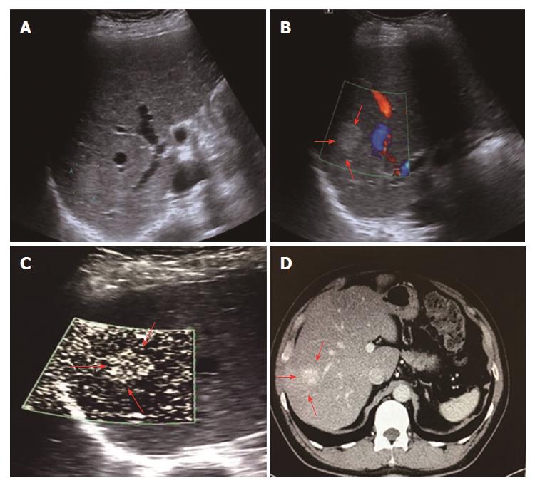
Figure 2 Diffuse dot-like type (type I) in a 52-year-old male diagnosed with hemangioma.
A: A high-echo lesion with a clear margin was evident in the right liver lobe; B: CDFI showed no blood flow signals for this lesion; C: SMI showed a diffuse dot-like microvascular structure; D: Contrast-enhanced CT showed diffuse enhancement of the lesion in the arterial phase. SMI: Superb microvascular imaging.
- Citation: He MN, Lv K, Jiang YX, Jiang TA. Application of superb microvascular imaging in focal liver lesions. World J Gastroenterol 2017; 23(43): 7765-7775
- URL: https://www.wjgnet.com/1007-9327/full/v23/i43/7765.htm
- DOI: https://dx.doi.org/10.3748/wjg.v23.i43.7765


