Copyright
©The Author(s) 2017.
World J Gastroenterol. Nov 21, 2017; 23(43): 7746-7755
Published online Nov 21, 2017. doi: 10.3748/wjg.v23.i43.7746
Published online Nov 21, 2017. doi: 10.3748/wjg.v23.i43.7746
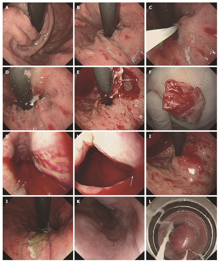
Figure 3 Glue adhesion to the endoscope in one patient.
A and B: Three GOV1 varices were observed; C: Obturation of one varix with tissue glue; D: The glue leaked out but the reversed endoscope blocked the vision; E: A large mass of solidified glue adhered to the endoscope and damaged the cardia; F: The glue was 1.5 cm in width and 3.0 cm in length; G and H: A large area of mucous was damaged in the esophagus and gastric cardia. Bleeding was observed, especially a gush of blood at the cardia; I: The bleeding at the cardia ceased after a 1% aethoxysklerol injection; J-L: The patient underwent follow-up 5 wk later. Extrusion of the glue from the gastric varices was observed, and EVL was performed to eradicate the esophageal varices. GOV: Gastroesophageal varices; EVL: Endoscopic variceal ligation.
- Citation: Guo YW, Miao HB, Wen ZF, Xuan JY, Zhou HX. Procedure-related complications in gastric variceal obturation with tissue glue. World J Gastroenterol 2017; 23(43): 7746-7755
- URL: https://www.wjgnet.com/1007-9327/full/v23/i43/7746.htm
- DOI: https://dx.doi.org/10.3748/wjg.v23.i43.7746


