Copyright
©The Author(s) 2017.
World J Gastroenterol. Nov 14, 2017; 23(42): 7531-7540
Published online Nov 14, 2017. doi: 10.3748/wjg.v23.i42.7531
Published online Nov 14, 2017. doi: 10.3748/wjg.v23.i42.7531
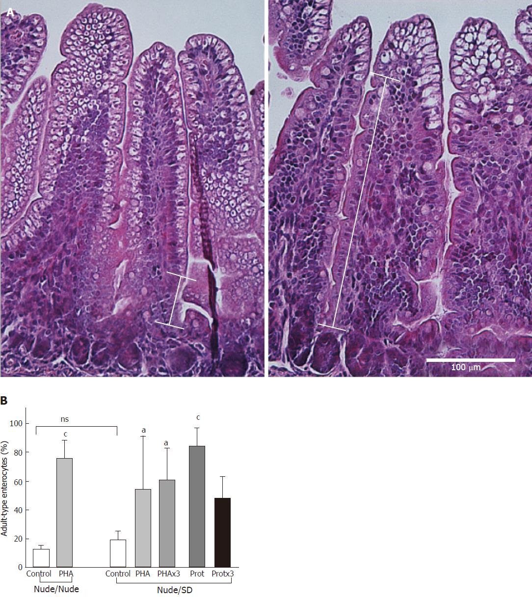
Figure 1 Epithelial maturation in the distal small intestine expressed as adult-type epithelium replacing the foetal-type vacuolated epithelium.
A: Photomicrograph (× 200, scale bar 100 μm) of H&E stained distal small intestine representative of a rat after PHA gavage (right) as compared to a control rat (left), with white bar connectors showing the portion of adult-type epithelium along the villus. B: Degree of intestinal epithelial maturation (%) in nude 14 day old rat pups treated by gavage with a kidney bean lectin - PHA (n = 4) or water (Control n = 3) and reared by their own mothers (Nude/Nude) and nude rat pups gavaged with PHA (n = 5) once or once a day for 3 d (PHAx3 n = 7), a protease once (Prot n = 4) or once a day for 3 days (Protx3 n = 7) or water (Control n = 5) and fostered by conventional SD dams (Nude/SD). Data presented as mean ± SD and differences were considered significant when P < 0.05. Significant differences between groups within nurturing groups (Nude/Nude or Nude/SD) indicated with aP < 0.05, bP < 0.01, cP < 0.001, dP < 0.0001 or non-significant (ns).
- Citation: Sureda EA, Gidlund C, Weström B, Prykhodko O. Induction of precocious intestinal maturation in T-cell deficient athymic neonatal rats. World J Gastroenterol 2017; 23(42): 7531-7540
- URL: https://www.wjgnet.com/1007-9327/full/v23/i42/7531.htm
- DOI: https://dx.doi.org/10.3748/wjg.v23.i42.7531


