Copyright
©The Author(s) 2017.
World J Gastroenterol. Oct 28, 2017; 23(40): 7337-7342
Published online Oct 28, 2017. doi: 10.3748/wjg.v23.i40.7337
Published online Oct 28, 2017. doi: 10.3748/wjg.v23.i40.7337
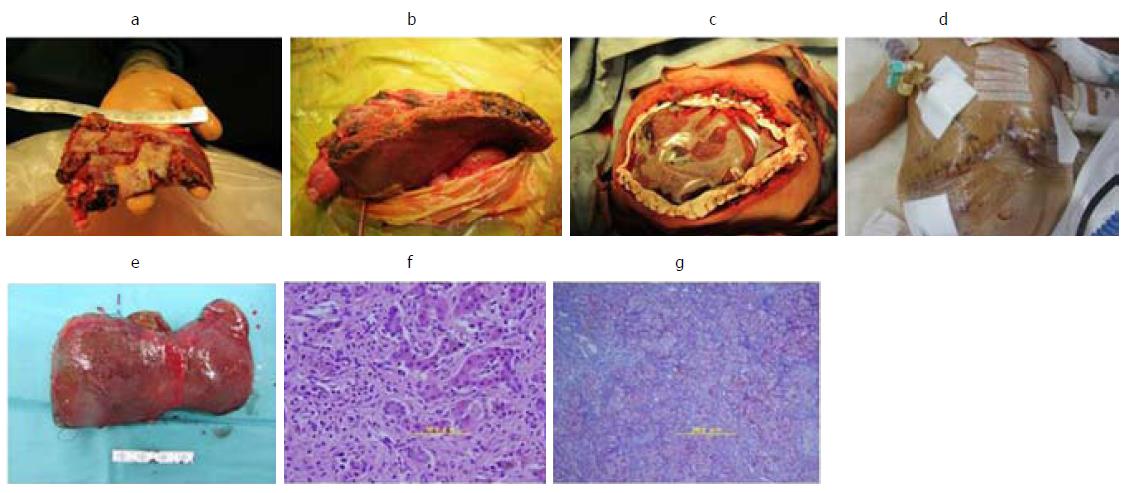
Figure 2 Images.
A: The S2 monosegment graft (107 g) on the back table; B: An image obtained after reperfusion, the graft was too large to close the abdominal fascia; C: The abdominal fascia could not be closed at the time of living donor liver transplantation (LDLT), excess water was removed by continuous hemodiafiltration after LDLT; D: Secondary skin closure was performed on postoperative day 5; E: The resected liver was 78 g; F: Hematoxylin and eosin staining revealed a marked lack of hepatocytes and the presence of multinucleated hepatocytes; G: Azan staining revealed widespread fibrosis around Glisson’s sheath and the parenchymal area (F3-4).
- Citation: Okada N, Sanada Y, Urahashi T, Ihara Y, Yamada N, Hirata Y, Katano T, Ushijima K, Otomo S, Fujita S, Mizuta K. Rescue case of low birth weight infant with acute hepatic failure. World J Gastroenterol 2017; 23(40): 7337-7342
- URL: https://www.wjgnet.com/1007-9327/full/v23/i40/7337.htm
- DOI: https://dx.doi.org/10.3748/wjg.v23.i40.7337


