Copyright
©The Author(s) 2017.
World J Gastroenterol. Oct 28, 2017; 23(40): 7253-7264
Published online Oct 28, 2017. doi: 10.3748/wjg.v23.i40.7253
Published online Oct 28, 2017. doi: 10.3748/wjg.v23.i40.7253
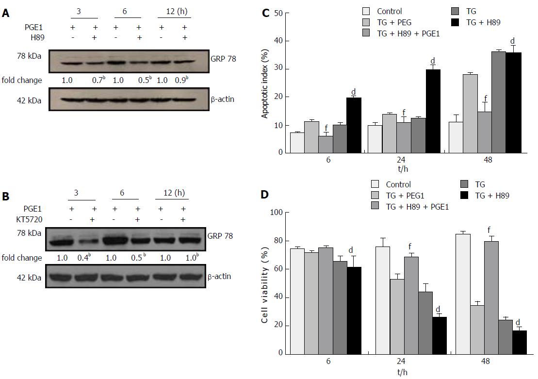
Figure 4 Prostaglandin E1 induced glucose-regulated protein 78 protein and mRNA expressions.
To determine GRP78 and CHOP protein or mRNA expressions, L02 cells and HepG2 cells were treated with 1 μmol/L PGE1 for 3, 6, 12 and 24 h; for detecting GADD34 and the p-PERK and p-eIF2α, L02 cells were treated with 1 μmol/L PGE1 for 6, 12, 24, 36 and 48 h. A: Expressions of GRP78, CHOP, GADD34, p-PERK and p-eIF2α in L02 cells were assessed by western blotting. One representative blot each from the three individual experiments is presented. The results of densitometric analysis are presented as a fold-change compared to those at 0 h (aP < 0.05, bP < 0.01); B: Expression of GRP78 in HepG2 cells was assessed by western blotting. One representative blot each from the three individual experiments is presented. The results of densitometric analysis are presented as a fold-change compared to those at 0 h (bP < 0.01); C: mRNA expression of GRP78 in L02 cells was assessed by quantitative real-time PCR (E). Histograms represent mean ± SD of three experiments (bP < 0.01 vs those at 0 h). CHOP: C/EBP homologous protein; ER: Endoplasmic reticulum; GADD34: Growth arrest and DNA damage-inducible gene 34; GRP78: Glucose-regulated protein 78; p-eIF2α: Phospho-eukaryotic translation initiation factor-2α; PGE1: Prostaglandin E1; p-PERK: Phospho-PKR-like ER kinase; TG: Thapsigargin.
- Citation: Yang FW, Fu Y, Li Y, He YH, Mu MY, Liu QC, Long J, Lin SD. Prostaglandin E1 protects hepatocytes against endoplasmic reticulum stress-induced apoptosis via protein kinase A-dependent induction of glucose-regulated protein 78 expression. World J Gastroenterol 2017; 23(40): 7253-7264
- URL: https://www.wjgnet.com/1007-9327/full/v23/i40/7253.htm
- DOI: https://dx.doi.org/10.3748/wjg.v23.i40.7253


