Copyright
©The Author(s) 2017.
World J Gastroenterol. Oct 28, 2017; 23(40): 7253-7264
Published online Oct 28, 2017. doi: 10.3748/wjg.v23.i40.7253
Published online Oct 28, 2017. doi: 10.3748/wjg.v23.i40.7253
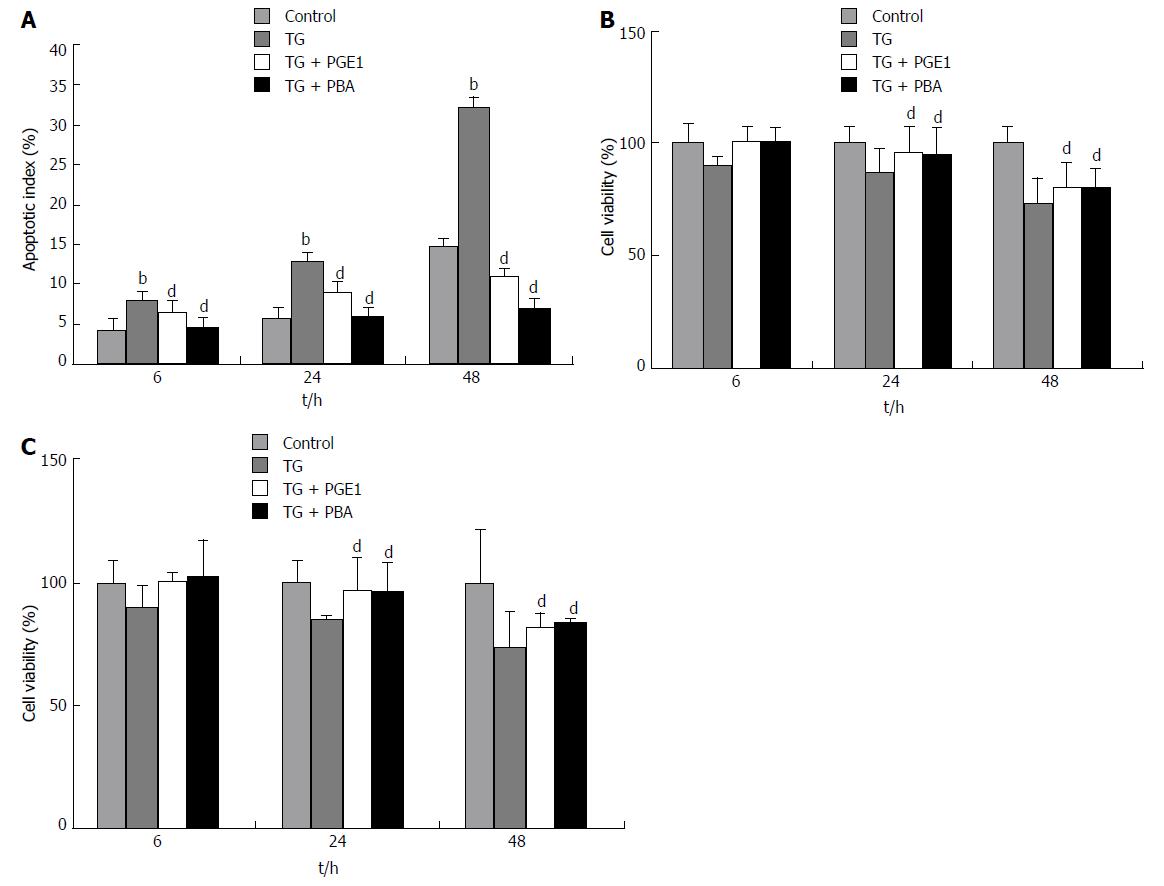
Figure 2 Prostaglandin E1 protected against thapsigargin-induced apoptosis in both L02 cells and HepG2 cells.
L02 cells and HepG2 cells were pretreated with PGE1 or PBA for 1 h and treated with a final concentration of 1 μmol/L TG for 6, 24 and 48 h. A: Apoptotic index of L02 cells was determined by flow cytometry. Histograms represent mean ± SD of five separate experiments, each of which was performed in triplicate. bP < 0.01 vs control at the same time point; dP < 0.01 vs TG at the same time point; B and C: Cell viability of L02 cells and HepG2 cells was determined by MTS assay. The absorbance was measured at 490 nm and cell viability was normalized as a percentage of control. Histograms represent mean ± SD of five separate experiments, each of which was performed in triplicate. dP < 0.01 vs TG at the same time point. MTS: [3-(4,5-dimethylthiazol-2-yl)-5-(3-carboxymethoxyphenyl)-2-(4-sulfophenyl)-2H-tetrazolium]; PBA: 4-phenylbutyric acid; PGE1: Prostaglandin E1; TG: Thapsigargin.
- Citation: Yang FW, Fu Y, Li Y, He YH, Mu MY, Liu QC, Long J, Lin SD. Prostaglandin E1 protects hepatocytes against endoplasmic reticulum stress-induced apoptosis via protein kinase A-dependent induction of glucose-regulated protein 78 expression. World J Gastroenterol 2017; 23(40): 7253-7264
- URL: https://www.wjgnet.com/1007-9327/full/v23/i40/7253.htm
- DOI: https://dx.doi.org/10.3748/wjg.v23.i40.7253


