Copyright
©The Author(s) 2017.
World J Gastroenterol. Jan 28, 2017; 23(4): 730-734
Published online Jan 28, 2017. doi: 10.3748/wjg.v23.i4.730
Published online Jan 28, 2017. doi: 10.3748/wjg.v23.i4.730
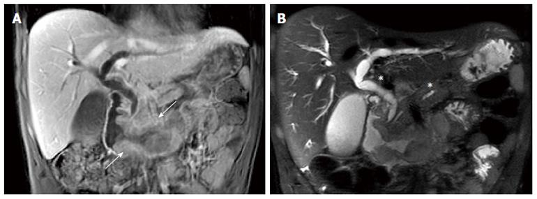
Figure 2 Magnetic resonance imaging.
A: MRI, coronal T1-weighted MR image with contrast showing the duodenal tissue thickening spreading from the second duodenum proximal part up to the duodenojejunal flexure (white arrow); B: MRI, coronal T2-weighted MR image showing the pancreatic duct and bile duct upstream swelling (white asterisks) without any secondary hepatic lesion. MRI: Magnetic resonance imaging.
- Citation: Barbieux J, Memeo R, De Blasi V, Suciu S, Faucher V, Averous G, Roy C, Marescaux J, Mutter D, Pessaux P. Real case of primitive embryonal duodenal carcinoma in a young man. World J Gastroenterol 2017; 23(4): 730-734
- URL: https://www.wjgnet.com/1007-9327/full/v23/i4/730.htm
- DOI: https://dx.doi.org/10.3748/wjg.v23.i4.730


