Copyright
©The Author(s) 2017.
World J Gastroenterol. Jan 28, 2017; 23(4): 573-589
Published online Jan 28, 2017. doi: 10.3748/wjg.v23.i4.573
Published online Jan 28, 2017. doi: 10.3748/wjg.v23.i4.573
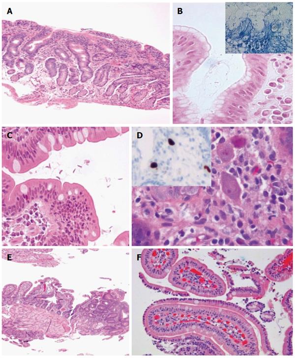
Figure 4 Gluten-sensitive enteropathy mimickers.
A: Lymphocytic gastritis with involvement of the duodenum (HE, × 100); B: H. pylori gastritis (inset, Giemsa staining) (HE and Giemsa × 630); C: Giardiasis (HE, × 400); D: Cytomegalovirus (CMV) infection (HE, × 6300) and inset showing anti-CMV antibody reacting against viral proteins using an avidin-biotin complex immunoperoxidase immunohistochemical detection (× 100); E: Focal adenomatous change in duodenum (HE, × 50); F: Sickle cell disease-related duodenitis (HE, × 200).
- Citation: Sergi C, Shen F, Bouma G. Intraepithelial lymphocytes, scores, mimickers and challenges in diagnosing gluten-sensitive enteropathy (celiac disease). World J Gastroenterol 2017; 23(4): 573-589
- URL: https://www.wjgnet.com/1007-9327/full/v23/i4/573.htm
- DOI: https://dx.doi.org/10.3748/wjg.v23.i4.573


