Copyright
©The Author(s) 2017.
World J Gastroenterol. Oct 21, 2017; 23(39): 7119-7128
Published online Oct 21, 2017. doi: 10.3748/wjg.v23.i39.7119
Published online Oct 21, 2017. doi: 10.3748/wjg.v23.i39.7119
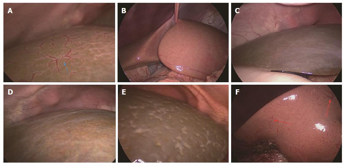
Figure 1 Laparoscopic images of the liver surface in the retrospective study.
A: HSST sign in a 70-day-old boy with biliary atresia (blue arrow); B: Image of a 64-day-old boy with Hirschsprung’s disease; C: Image of an 82-day-old boy with infantile hepatitis; D: Image of a 72-day-old girl with biliary hypoplasia; E: Image of a 70-day-old boy with total parenteral nutrition-induced cholestatic cirrhosis; F: Small vessel plexuses (red arrows) observed in a 55-day-old boy with cytomegalovirus hepatitis. The HSST sign does not exist in the images B-E. HSST: Hepatic subcapsular spider-like telangiectasis.
- Citation: Zhou Y, Jiang M, Tang ST, Yang L, Zhang X, Yang DH, Xiong M, Li S, Cao GQ, Wang Y. Laparoscopic finding of a hepatic subcapsular spider-like telangiectasis sign in biliary atresia. World J Gastroenterol 2017; 23(39): 7119-7128
- URL: https://www.wjgnet.com/1007-9327/full/v23/i39/7119.htm
- DOI: https://dx.doi.org/10.3748/wjg.v23.i39.7119


