Copyright
©The Author(s) 2017.
World J Gastroenterol. Oct 14, 2017; 23(38): 7054-7058
Published online Oct 14, 2017. doi: 10.3748/wjg.v23.i38.7054
Published online Oct 14, 2017. doi: 10.3748/wjg.v23.i38.7054
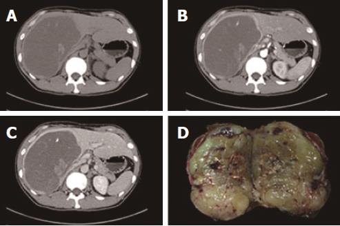
Figure 1 Computed tomography imaging and surgical specimen.
Dynamic contrast-enhanced computed tomography (CT) imaging showed that a cyst-like lesion (A) contained a stripe-like, slightly hyperdense area and patchy hypodense areas that were hyper-enhanced in the arterial phase (B) and hypo-enhanced in the portal phase (C), whereas the central portion remained unenhanced throughout the arterial and portal phases. Sections through the tumor disclosed grayish-yellow, solid tissue that showed relatively homogenous internal structures (D).
- Citation: Wen J, Zhao W, Li C, Shen JY, Wen TF. High-grade myofibroblastic sarcoma in the liver: A case report. World J Gastroenterol 2017; 23(38): 7054-7058
- URL: https://www.wjgnet.com/1007-9327/full/v23/i38/7054.htm
- DOI: https://dx.doi.org/10.3748/wjg.v23.i38.7054


