Copyright
©The Author(s) 2017.
World J Gastroenterol. Oct 14, 2017; 23(38): 6942-6951
Published online Oct 14, 2017. doi: 10.3748/wjg.v23.i38.6942
Published online Oct 14, 2017. doi: 10.3748/wjg.v23.i38.6942
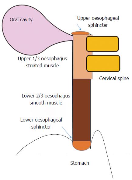
Figure 1 Anatomy of the oesophagus.
Disease of the upper 1/3 of the oesophagus causing dysphagia may include extrinsic compression (e.g., cervical osteophytes), or dysfunction secondary to rheumatological conditions (e.g., Sjogrens’s syndrome) or in eosinophilic oesophagitis (along with the lower oesophagus). The lower oesophagus can be afflicted in scleroderma, gastroesophageal reflux disease and in eosinophilic oesophagitis.
- Citation: Philpott H, Garg M, Tomic D, Balasubramanian S, Sweis R. Dysphagia: Thinking outside the box. World J Gastroenterol 2017; 23(38): 6942-6951
- URL: https://www.wjgnet.com/1007-9327/full/v23/i38/6942.htm
- DOI: https://dx.doi.org/10.3748/wjg.v23.i38.6942


