Copyright
©The Author(s) 2017.
World J Gastroenterol. Oct 7, 2017; 23(37): 6911-6919
Published online Oct 7, 2017. doi: 10.3748/wjg.v23.i37.6911
Published online Oct 7, 2017. doi: 10.3748/wjg.v23.i37.6911
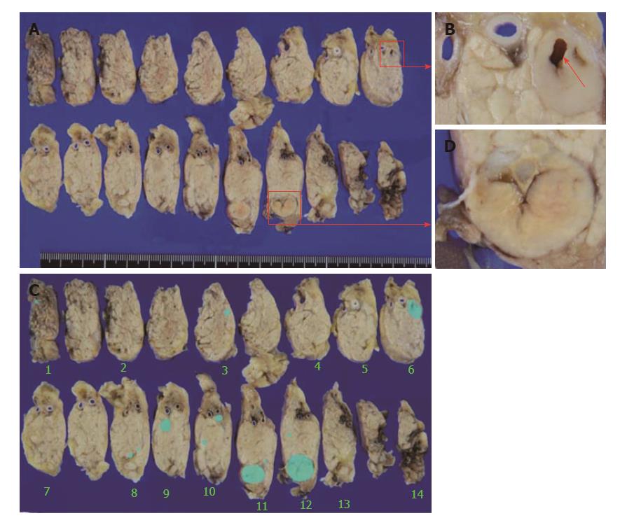
Figure 5 Macroscopic findings for the resected pancreas.
A: The main cystic lesion has morphologically changed to a 13-mm nodule (red square C). The microtumor in the pancreatic body on which EUS-FNA was performed was separate from the main lesion; B: A scar from EUS-FNA is evident in the microtumor (red arrow); C: The main nodule shows internal scarring; D: The main lesion and two microtumors detected on EUS, and another 11 new microtumors (1-3 mm) (blue areas). EUS-FNA: Endoscopic ultrasound-fine needle aspiration.
- Citation: Sagami R, Nishikiori H, Ikuyama S, Murakami K. Rupture of small cystic pancreatic neuroendocrine tumor with many microtumors. World J Gastroenterol 2017; 23(37): 6911-6919
- URL: https://www.wjgnet.com/1007-9327/full/v23/i37/6911.htm
- DOI: https://dx.doi.org/10.3748/wjg.v23.i37.6911


