Copyright
©The Author(s) 2017.
World J Gastroenterol. Oct 7, 2017; 23(37): 6894-6901
Published online Oct 7, 2017. doi: 10.3748/wjg.v23.i37.6894
Published online Oct 7, 2017. doi: 10.3748/wjg.v23.i37.6894
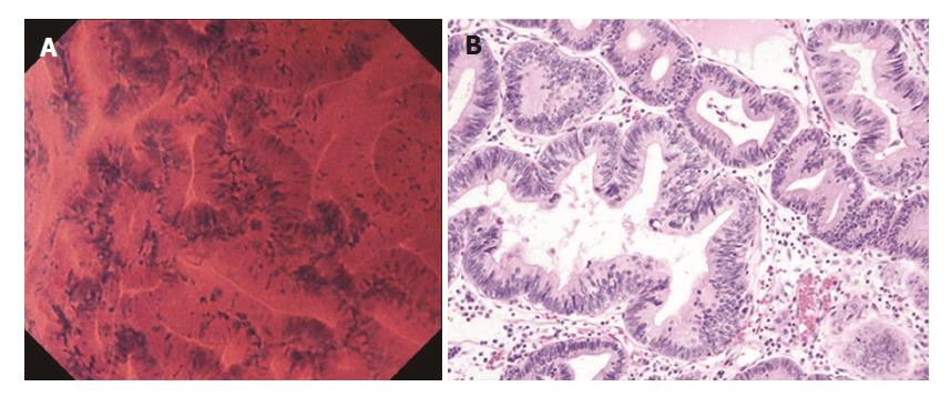
Figure 4 Well differentiated adenocarcinoma.
A: Endocytoscopic appearance of a well differentiated adenocarcinoma. The glands were branched irregularly and the width of the lumen varied. The epithelium was arranged in a disorderly fashion. The nuclei were deeply stained and pseudostratified; B: Histological appearance of the corresponding well-differentiated adenocarcinoma (H&E-stained horizontal section, × 200).
- Citation: Tsurudome I, Miyahara R, Funasaka K, Furukawa K, Matsushita M, Yamamura T, Ishikawa T, Ohno E, Nakamura M, Kawashima H, Watanabe O, Nakaguro M, Satou A, Hirooka Y, Goto H. In vivo histological diagnosis for gastric cancer using endocytoscopy. World J Gastroenterol 2017; 23(37): 6894-6901
- URL: https://www.wjgnet.com/1007-9327/full/v23/i37/6894.htm
- DOI: https://dx.doi.org/10.3748/wjg.v23.i37.6894


