Copyright
©The Author(s) 2017.
World J Gastroenterol. Oct 7, 2017; 23(37): 6833-6844
Published online Oct 7, 2017. doi: 10.3748/wjg.v23.i37.6833
Published online Oct 7, 2017. doi: 10.3748/wjg.v23.i37.6833
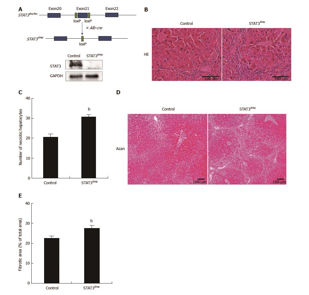
Figure 1 Establishment of hepatic-STAT3 deficient (STAT3Δhep) mice.
Hepatic-STAT3 deficiency enhanced thioacetamide-induced liver injury. A: The exon 21 of STAT3 was flanked by two identically oriented loxP sites (STAT3flox/flox). To disrupt hepatic-STAT3 gene, STAT3flox/flox mice were crossed with mice carrying albumin promoter-driven Cre transgene (Alb-Cre). Deficiency of hepatic-STAT3 protein was confirmed by immunoblotting analysis. B: Representative hematoxylin and eosin (H&E) staining images of control and STAT3Δhep livers with TAA treatment for 16 wk. Hepatic-STAT3 deficiency accelerated inflammation cells infiltration and hepatocytes necrosis. C: Quantification of necrotic hepatocytes. Necrotic hepatocyte was identified with the eosinophilic/vacuolated cytoplasm and nuclear destruction such as karyolysis or Pyknosis. Values represent means ± SE of the mean. bP < 0.01 (P = 0.000269) vs control. D: Representative Azan staining of control and STAT3Δhep livers with TAA treatment for 16 wk. E: Quantification of the ratio of fibrosis area to the total area in control and STAT3Δhep livers. Values represent means ± SE of the mean. bP < 0.01 (P = 0.00235) vs control. TAA: Thioacetamide.
- Citation: Abe M, Yoshida T, Akiba J, Ikezono Y, Wada F, Masuda A, Sakaue T, Tanaka T, Iwamoto H, Nakamura T, Sata M, Koga H, Yoshimura A, Torimura T. STAT3 deficiency prevents hepatocarcinogenesis and promotes biliary proliferation in thioacetamide-induced liver injury. World J Gastroenterol 2017; 23(37): 6833-6844
- URL: https://www.wjgnet.com/1007-9327/full/v23/i37/6833.htm
- DOI: https://dx.doi.org/10.3748/wjg.v23.i37.6833


