Copyright
©The Author(s) 2017.
World J Gastroenterol. Sep 21, 2017; 23(35): 6467-6473
Published online Sep 21, 2017. doi: 10.3748/wjg.v23.i35.6467
Published online Sep 21, 2017. doi: 10.3748/wjg.v23.i35.6467
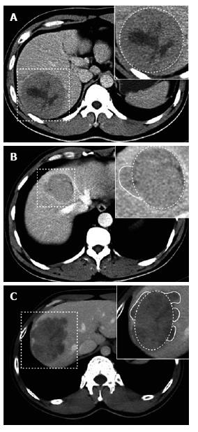
Figure 1 Tumor margins on the liver computed tomography map.
A: Portovenous phase CT showing smooth margin at liver segment VI in a 36-year-old male patient without postoperative recurrence (dashed box); B: Portovenous phase CT showing focal extranodular type at liver segments V and VIII in a 38-year-old female patient with postoperative recurrence (dashed box); C: Portovenous phase CT showing multinodular type at liver segments V and VI in a 35-year-old male patient with postoperative recurrence (dashed box). CT: Computed tomography.
- Citation: Zhang W, Lai SL, Chen J, Xie D, Wu FX, Jin GQ, Su DK. Validated preoperative computed tomography risk estimation for postoperative hepatocellular carcinoma recurrence. World J Gastroenterol 2017; 23(35): 6467-6473
- URL: https://www.wjgnet.com/1007-9327/full/v23/i35/6467.htm
- DOI: https://dx.doi.org/10.3748/wjg.v23.i35.6467


