Copyright
©The Author(s) 2017.
World J Gastroenterol. Sep 21, 2017; 23(35): 6437-6447
Published online Sep 21, 2017. doi: 10.3748/wjg.v23.i35.6437
Published online Sep 21, 2017. doi: 10.3748/wjg.v23.i35.6437
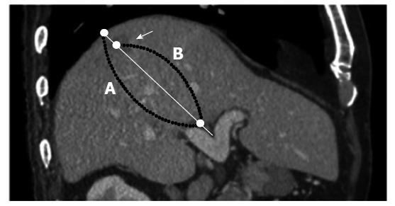
Figure 1 A 73-year-old man with hepatocellular carcinoma.
An MPR image of the arterial phase shows a well-defined enhanced mass in segment 7 (arrowhead) and the bifurcation of the right and left branches of the portal vein. HCC location coefficient = distance from the medial surface of liver to the central HCC tumor (B)/diameter of the liver (A). MPR: Multi-planar reconstructed images; HCC: Hepatocellular carcinoma.
- Citation: Miki I, Murata S, Uchiyama F, Yasui D, Ueda T, Sugihara F, Saito H, Yamaguchi H, Murakami R, Kawamoto C, Uchida E, Kumita SI. Evaluation of the relationship between hepatocellular carcinoma location and transarterial chemoembolization efficacy. World J Gastroenterol 2017; 23(35): 6437-6447
- URL: https://www.wjgnet.com/1007-9327/full/v23/i35/6437.htm
- DOI: https://dx.doi.org/10.3748/wjg.v23.i35.6437


