Copyright
©The Author(s) 2017.
World J Gastroenterol. Sep 14, 2017; 23(34): 6201-6211
Published online Sep 14, 2017. doi: 10.3748/wjg.v23.i34.6201
Published online Sep 14, 2017. doi: 10.3748/wjg.v23.i34.6201
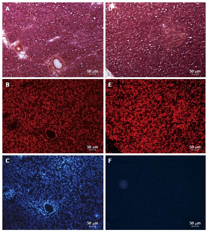
Figure 6 Fluorescence microscopy image of cryosections from left lobe of the pancreas showing distribution of endocrine and exocrine cells with nuclei stained in disto-medial (A-C) and proximo-lateral (D-F) portions.
A and D: Serial sections stained with hematoxyline-eosine; B-E: Serial sections stained with the nuclear marker TO-PRO®3 iodide; C and F: Serial sections showing the location the nuclear dye Hoechst (Bizbenzimida H 33258 fluorochrome).
- Citation: Latorre R, López-Albors O, Soria F, Morcillo E, Esteban P, Pérez-Cuadrado-Robles E, Pérez-Cuadrado-Martínez E. Evidences supporting the vascular etiology of post-double balloon enteroscopy pancreatitis: Study in porcine model. World J Gastroenterol 2017; 23(34): 6201-6211
- URL: https://www.wjgnet.com/1007-9327/full/v23/i34/6201.htm
- DOI: https://dx.doi.org/10.3748/wjg.v23.i34.6201


