Copyright
©The Author(s) 2017.
World J Gastroenterol. Sep 14, 2017; 23(34): 6201-6211
Published online Sep 14, 2017. doi: 10.3748/wjg.v23.i34.6201
Published online Sep 14, 2017. doi: 10.3748/wjg.v23.i34.6201
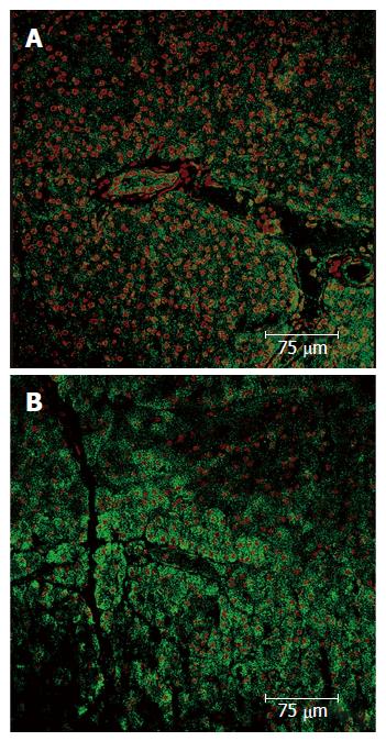
Figure 2 Pancreas immunohistochemistry.
A: Expression of the VEGF in a normal parenchyma; B: Pancreatic acini with normal structure and nuclei, which express VEGF. In upper right corner a ischemic zone where less VEGF expression is shown, the pancreatic acini structure has been lost and nuclei are pyknotic. VEGF: Vascular endothelial growth factor.
- Citation: Latorre R, López-Albors O, Soria F, Morcillo E, Esteban P, Pérez-Cuadrado-Robles E, Pérez-Cuadrado-Martínez E. Evidences supporting the vascular etiology of post-double balloon enteroscopy pancreatitis: Study in porcine model. World J Gastroenterol 2017; 23(34): 6201-6211
- URL: https://www.wjgnet.com/1007-9327/full/v23/i34/6201.htm
- DOI: https://dx.doi.org/10.3748/wjg.v23.i34.6201


