Copyright
©The Author(s) 2017.
World J Gastroenterol. Sep 7, 2017; 23(33): 6147-6154
Published online Sep 7, 2017. doi: 10.3748/wjg.v23.i33.6147
Published online Sep 7, 2017. doi: 10.3748/wjg.v23.i33.6147
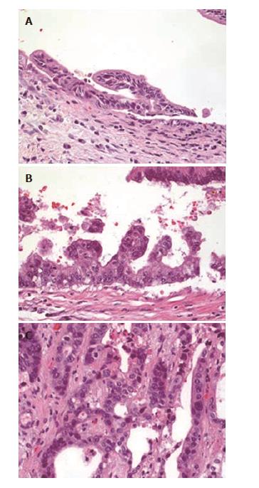
Figure 2 Histology from explanted livers diagnosed with biliary neoplasia.
A: Bile duct from explanted liver with low-grade dysplasia; B: Bile duct from explanted liver with high-grade dysplasia; C: Cholangiocarcinoma from explanted liver.
- Citation: Boyd S, Vannas M, Jokelainen K, Isoniemi H, Mäkisalo H, Färkkilä MA, Arola J. Suspicious brush cytology is an indication for liver transplantation evaluation in primary sclerosing cholangitis. World J Gastroenterol 2017; 23(33): 6147-6154
- URL: https://www.wjgnet.com/1007-9327/full/v23/i33/6147.htm
- DOI: https://dx.doi.org/10.3748/wjg.v23.i33.6147


