Copyright
©The Author(s) 2017.
World J Gastroenterol. Aug 28, 2017; 23(32): 5936-5944
Published online Aug 28, 2017. doi: 10.3748/wjg.v23.i32.5936
Published online Aug 28, 2017. doi: 10.3748/wjg.v23.i32.5936
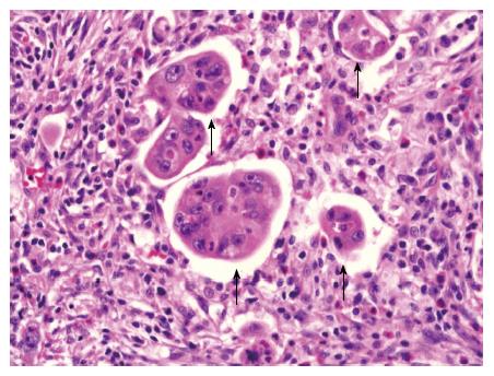
Figure 2 Representative histopathological micrograph of poorly differentiated clusters (magnification × 400).
Hematoxylin and eosin stain of tumor section showing cancer cell clusters of ≥ 5 carcinoma cells lacking a glandular formation (poorly differentiated clusters, black arrows).
- Citation: Yim K, Won DD, Lee IK, Oh ST, Jung ES, Lee SH. Novel predictors for lymph node metastasis in submucosal invasive colorectal carcinoma. World J Gastroenterol 2017; 23(32): 5936-5944
- URL: https://www.wjgnet.com/1007-9327/full/v23/i32/5936.htm
- DOI: https://dx.doi.org/10.3748/wjg.v23.i32.5936


