Copyright
©The Author(s) 2017.
World J Gastroenterol. Aug 28, 2017; 23(32): 5925-5935
Published online Aug 28, 2017. doi: 10.3748/wjg.v23.i32.5925
Published online Aug 28, 2017. doi: 10.3748/wjg.v23.i32.5925
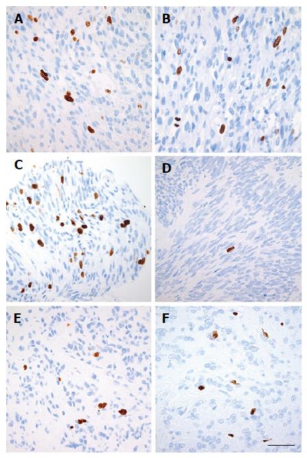
Figure 4 Ki-67-immunostaining of gastrointestinal stromal tumors-tumor tissue in three endoscopic ultrasound-biopsies and in the three corresponding resected specimens.
Digital photos (magnification × 40): EUS-biopsy-tissue (left) and resected specimen tissue (right). Cell nuclei (brown color) are positive for Ki-67 while other cell nuclei (blue color) are negative for Ki-67. Scale bar equals 50 μm. A and B: Case #18, Neo- group (KIT exon 11 D579del, Ki-67EUS: 6.6%, Ki-67SURG: 6.3%); C and D: Case #22, Neo + s group (KIT exon 11 V559D, Ki-67EUS: 5.6%, Ki-67SURG 0.3%); E and F: Case #26, Neo + r group (PDGFRA exon 18 p.D842V, Ki-67EUS: 2.7%, Ki-67SURG 2.5%). EUS: Endoscopic ultrasound.
- Citation: Hedenström P, Nilsson B, Demir A, Andersson C, Enlund F, Nilsson O, Sadik R. Characterizing gastrointestinal stromal tumors and evaluating neoadjuvant imatinib by sequencing of endoscopic ultrasound-biopsies. World J Gastroenterol 2017; 23(32): 5925-5935
- URL: https://www.wjgnet.com/1007-9327/full/v23/i32/5925.htm
- DOI: https://dx.doi.org/10.3748/wjg.v23.i32.5925


