Copyright
©The Author(s) 2017.
World J Gastroenterol. Aug 28, 2017; 23(32): 5849-5859
Published online Aug 28, 2017. doi: 10.3748/wjg.v23.i32.5849
Published online Aug 28, 2017. doi: 10.3748/wjg.v23.i32.5849
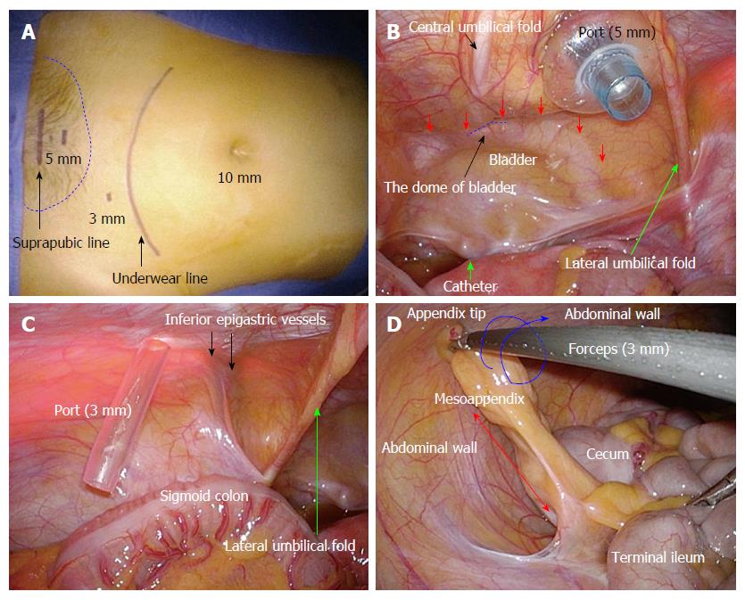
Figure 2 Major techniques during laparoscopic appendectomy.
A: A suprapubic port (5 mm) for a flexible laparoscope is placed within the area of pubic hair (dotted blue line) to hide the postoperative stab scar. A left lateral port (3 mm) is placed as low as possible, to enable an adequate angle for the working forceps and to hide the postoperative stab scar by underwear; B: The bladder wall (red arrows), the dome of the bladder (dotted blue line), and the central umbilical fold should be recognized. Although the suprapubic peritoneum easily extends during port insertion, a suprapubic port should be placed without bladder injury; C: Any injury of the left inferior epigastric vessels should be avoided; D: Countertraction of the mesoappendix (red arrow) should be made without obstruction of the abdominal wall. Gripping and rotating forces of 3-mm forceps are sufficient. The appendix can be shortened in a rolled-in fashion (blue arrow) to avoid any disturbance by the abdominal wall.
- Citation: Hori T, Machimoto T, Kadokawa Y, Hata T, Ito T, Kato S, Yasukawa D, Aisu Y, Kimura Y, Sasaki M, Takamatsu Y, Kitano T, Hisamori S, Yoshimura T. Laparoscopic appendectomy for acute appendicitis: How to discourage surgeons using inadequate therapy. World J Gastroenterol 2017; 23(32): 5849-5859
- URL: https://www.wjgnet.com/1007-9327/full/v23/i32/5849.htm
- DOI: https://dx.doi.org/10.3748/wjg.v23.i32.5849


