Copyright
©The Author(s) 2017.
World J Gastroenterol. Aug 21, 2017; 23(31): 5817-5822
Published online Aug 21, 2017. doi: 10.3748/wjg.v23.i31.5817
Published online Aug 21, 2017. doi: 10.3748/wjg.v23.i31.5817
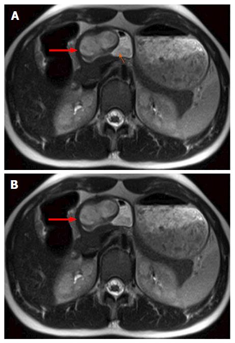
Figure 1 Magnetic resonance imaging findings for case 1, using transverse T2-and T1-weighted magnetic resonance imaging.
A: Transverse T2-weighted magnetic resonance imaging (MRI) showing well circumscribed, endophytic mass (thick red arrow) of the gastric antrum which is partially filled with fluid (thin orange arrow); B: T1-weighted MRI after contrast media application showing homogeneous wall enhancement of the well circumscribed mass (thick red arrow).
- Citation: Szurian K, Till H, Amerstorfer E, Hinteregger N, Mischinger HJ, Liegl-Atzwanger B, Brcic I. Rarity among benign gastric tumors: Plexiform fibromyxoma - Report of two cases. World J Gastroenterol 2017; 23(31): 5817-5822
- URL: https://www.wjgnet.com/1007-9327/full/v23/i31/5817.htm
- DOI: https://dx.doi.org/10.3748/wjg.v23.i31.5817


