Copyright
©The Author(s) 2017.
World J Gastroenterol. Aug 21, 2017; 23(31): 5798-5808
Published online Aug 21, 2017. doi: 10.3748/wjg.v23.i31.5798
Published online Aug 21, 2017. doi: 10.3748/wjg.v23.i31.5798
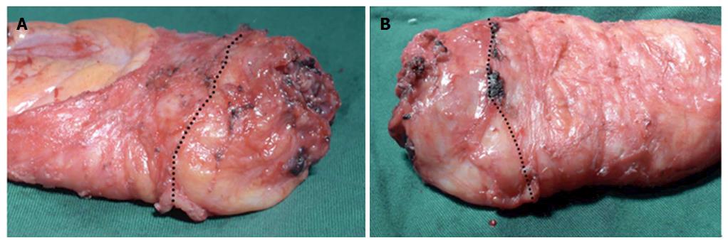
Figure 3 Specimen was examined by a pathologist.
A: The anterior side of specimen; B: The posterior side of specimen. The black dotted line shows the boundary of transabdominal total mesorectal excision (TME) and retrograde transanal TME. The lower rectum had a smoother mesorectum surface compared with the upper rectum. TME: Total mesorectal excision.
- Citation: Xu C, Song HY, Han SL, Ni SC, Zhang HX, Xing CG. Simple instruments facilitating achievement of transanal total mesorectal excision in male patients. World J Gastroenterol 2017; 23(31): 5798-5808
- URL: https://www.wjgnet.com/1007-9327/full/v23/i31/5798.htm
- DOI: https://dx.doi.org/10.3748/wjg.v23.i31.5798


