Copyright
©The Author(s) 2017.
World J Gastroenterol. Aug 21, 2017; 23(31): 5787-5797
Published online Aug 21, 2017. doi: 10.3748/wjg.v23.i31.5787
Published online Aug 21, 2017. doi: 10.3748/wjg.v23.i31.5787
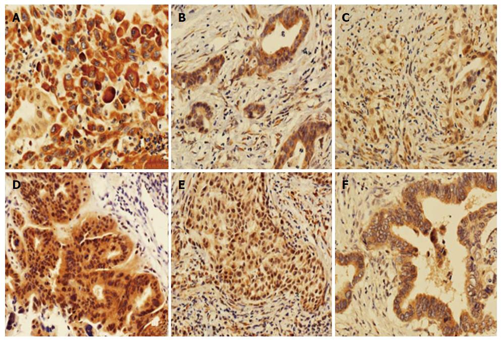
Figure 2 Immunohistochemical staining of tumor tissues (magnification, × 200).
A-C: Cytoplasm staining with 3+, 2+ and 1+ intensity, respectively; D-F: Nuclear staining with 2+, 1+ and 0+ intensity, respectively.
- Citation: Xie Y, Lin JZ, Wang AQ, Xu WY, Long JY, Luo YF, Shi J, Liang ZY, Sang XT, Zhao HT. Threonine and tyrosine kinase may serve as a prognostic biomarker for gallbladder cancer. World J Gastroenterol 2017; 23(31): 5787-5797
- URL: https://www.wjgnet.com/1007-9327/full/v23/i31/5787.htm
- DOI: https://dx.doi.org/10.3748/wjg.v23.i31.5787


