Copyright
©The Author(s) 2017.
World J Gastroenterol. Aug 14, 2017; 23(30): 5619-5633
Published online Aug 14, 2017. doi: 10.3748/wjg.v23.i30.5619
Published online Aug 14, 2017. doi: 10.3748/wjg.v23.i30.5619
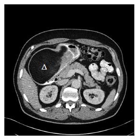
Figure 2 Abdominal computerized tomography findings in patient 1.
A 63-year-old male (patient 1) presented with acute melena and hemoglobin decline to 6.2 g/dL, and esophagogastroduodenoscopy revealed a huge, submucosal mass with a smooth overlying surface and exhibiting the pillow sign characteristic of a submucosal lipoma. Illustrated abdominal computerized tomography shows an approximately 13.4 cm × 8.2 cm × 8.4 cm mostly homogeneous, hypodense mass with a characteristic attenuation of fat (-90.2 Hounsfield units) extending from proximal gastric body through entire antrum. The normal-appearing very proximal stomach is filled with oral contrast without a mass, and leads to a very narrow, compressed, distal and dorsal, gastric channel containing oral contrast that passes into the duodenum. Triangle: antral giant gastric lipoma which has the characteristic hypodense attenuation of fat.
- Citation: Cappell MS, Stevens CE, Amin M. Systematic review of giant gastric lipomas reported since 1980 and report of two new cases in a review of 117110 esophagogastroduodenoscopies. World J Gastroenterol 2017; 23(30): 5619-5633
- URL: https://www.wjgnet.com/1007-9327/full/v23/i30/5619.htm
- DOI: https://dx.doi.org/10.3748/wjg.v23.i30.5619


