Copyright
©The Author(s) 2017.
World J Gastroenterol. Aug 14, 2017; 23(30): 5567-5578
Published online Aug 14, 2017. doi: 10.3748/wjg.v23.i30.5567
Published online Aug 14, 2017. doi: 10.3748/wjg.v23.i30.5567
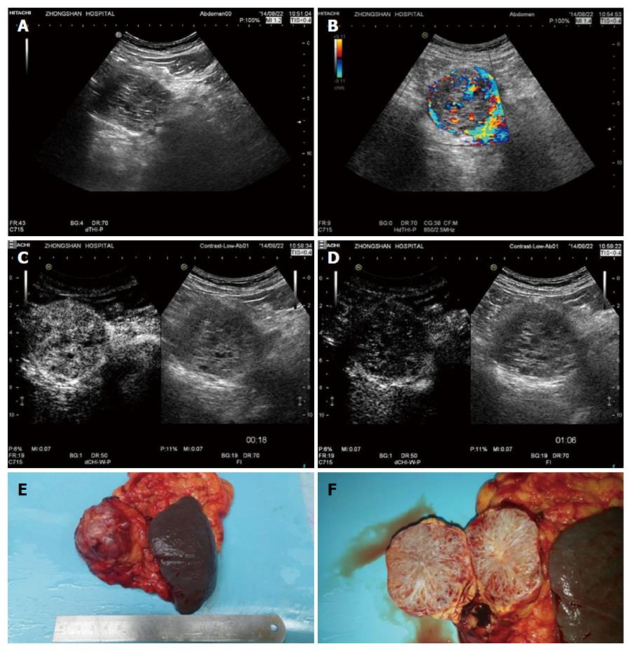
Figure 5 Histopathologically proven serous microcystic serous pancreatic neoplasia.
A: A solid-cystic lesion was detected in the head of pancreas with B-mode ultrasound; B: Multiple interlesional color flow signals were detected using colour Doppler imaging; C: Contrast enhanced ultrasound showed the lesion to hyperenhance in the arterial phase; D: Isoenhance in the late phase; E and F: Surgical pathology shows the typical honeycomb structure.
- Citation: Dietrich CF, Dong Y, Jenssen C, Ciaravino V, Hocke M, Wang WP, Burmester E, Moeller K, Atkinson NS, Capelli P, D’Onofrio M. Serous pancreatic neoplasia, data and review. World J Gastroenterol 2017; 23(30): 5567-5578
- URL: https://www.wjgnet.com/1007-9327/full/v23/i30/5567.htm
- DOI: https://dx.doi.org/10.3748/wjg.v23.i30.5567


