Copyright
©The Author(s) 2017.
World J Gastroenterol. Aug 14, 2017; 23(30): 5460-5468
Published online Aug 14, 2017. doi: 10.3748/wjg.v23.i30.5460
Published online Aug 14, 2017. doi: 10.3748/wjg.v23.i30.5460
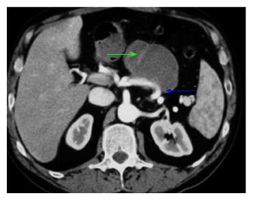
Figure 1 Computed tomography arterial and venous phase showing a pseudocyst (green arrows) eroding the splenic artery (blue arrows)[90].
- Citation: Evans RP, Mourad MM, Pall G, Fisher SG, Bramhall SR. Pancreatitis: Preventing catastrophic haemorrhage. World J Gastroenterol 2017; 23(30): 5460-5468
- URL: https://www.wjgnet.com/1007-9327/full/v23/i30/5460.htm
- DOI: https://dx.doi.org/10.3748/wjg.v23.i30.5460


