Copyright
©The Author(s) 2017.
World J Gastroenterol. Aug 7, 2017; 23(29): 5422-5430
Published online Aug 7, 2017. doi: 10.3748/wjg.v23.i29.5422
Published online Aug 7, 2017. doi: 10.3748/wjg.v23.i29.5422
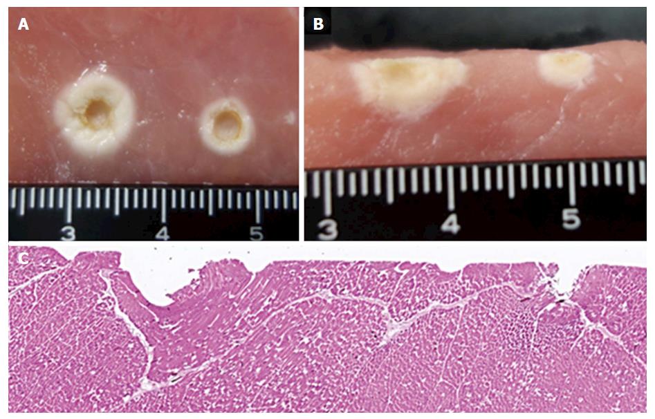
Figure 5 Macroscopic view after coagulation by the ball electrode with a 3-mm tip and microscopic view after coagulation by the FlushKnife-BT in the porcine block.
A: Macroscopic view of the front surface after coagulation using the ball electrode with a 3-mm tip. The coagulation at the left and right is the result of the F1-10 and S methods, respectively; B: Macroscopic view of a transverse section after coagulation using the ball electrode with a 3-mm tip; C: Microscopic view of the hematoxylin-eosin staining after coagulation by the FlushKnife-BT 2.5 mm. The coagulation at the left and right is the result of the F1-10 and S methods, respectively.
- Citation: Ishida T, Toyonaga T, Ohara Y, Nakashige T, Kitamura Y, Ariyoshi R, Takihara H, Baba S, Yoshizaki T, Kawara F, Tanaka S, Morita Y, Umegaki E, Hoshi N, Azuma T. Efficacy of forced coagulation with low high-frequency power setting during endoscopic submucosal dissection. World J Gastroenterol 2017; 23(29): 5422-5430
- URL: https://www.wjgnet.com/1007-9327/full/v23/i29/5422.htm
- DOI: https://dx.doi.org/10.3748/wjg.v23.i29.5422


