Copyright
©The Author(s) 2017.
World J Gastroenterol. Aug 7, 2017; 23(29): 5282-5294
Published online Aug 7, 2017. doi: 10.3748/wjg.v23.i29.5282
Published online Aug 7, 2017. doi: 10.3748/wjg.v23.i29.5282
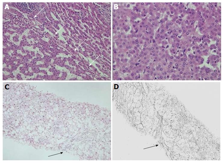
Figure 3 Well differentiated, (grade 1) hepatocellular carcinoma and early hepatocellular carcinoma with diffuse fatty change.
A: White arrows indicate the interface between HCC (left) and background liver (right); B: HCC cells show high nuclear/cytoplasmic ratio and minimal nuclear atypia. A: H/E × 100, B: H/E × 200; C and D: Early HCC with diffuse fatty change. Black arrowhead depicts a preserved portal tract. Gomori stain shows rarefaction of reticulin network. C: H/E × 100, D: Gomori stain × 100. HCC: Hepatocellular carcinoma.
- Citation: Dimitroulis D, Damaskos C, Valsami S, Davakis S, Garmpis N, Spartalis E, Athanasiou A, Moris D, Sakellariou S, Kykalos S, Tsourouflis G, Garmpi A, Delladetsima I, Kontzoglou K, Kouraklis G. From diagnosis to treatment of hepatocellular carcinoma: An epidemic problem for both developed and developing world. World J Gastroenterol 2017; 23(29): 5282-5294
- URL: https://www.wjgnet.com/1007-9327/full/v23/i29/5282.htm
- DOI: https://dx.doi.org/10.3748/wjg.v23.i29.5282


