Copyright
©The Author(s) 2017.
World J Gastroenterol. Aug 7, 2017; 23(29): 5282-5294
Published online Aug 7, 2017. doi: 10.3748/wjg.v23.i29.5282
Published online Aug 7, 2017. doi: 10.3748/wjg.v23.i29.5282
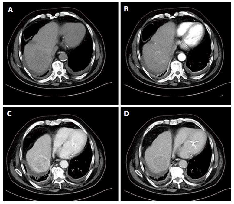
Figure 2 Multiphasic computed tomography in a large hepatocellular carcinoma located in the right liver lobe.
A: Unenhanced image; B: Lesion’s enhancement in the late hepatic arterial phase; C: Lesion’s “washout” in the portal venous phase; D: Delayed phase image. The lesion has capsule appearance most shown in the portal venous and delayed phase.
- Citation: Dimitroulis D, Damaskos C, Valsami S, Davakis S, Garmpis N, Spartalis E, Athanasiou A, Moris D, Sakellariou S, Kykalos S, Tsourouflis G, Garmpi A, Delladetsima I, Kontzoglou K, Kouraklis G. From diagnosis to treatment of hepatocellular carcinoma: An epidemic problem for both developed and developing world. World J Gastroenterol 2017; 23(29): 5282-5294
- URL: https://www.wjgnet.com/1007-9327/full/v23/i29/5282.htm
- DOI: https://dx.doi.org/10.3748/wjg.v23.i29.5282


