Copyright
©The Author(s) 2017.
World J Gastroenterol. Jul 28, 2017; 23(28): 5115-5126
Published online Jul 28, 2017. doi: 10.3748/wjg.v23.i28.5115
Published online Jul 28, 2017. doi: 10.3748/wjg.v23.i28.5115
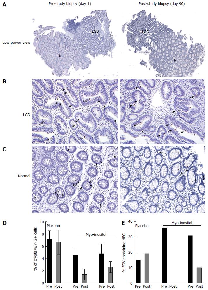
Figure 4 Myo-inositol reduces the number of pβ-cat-positive cells in ulcerative colitis patients.
A: Representative low power images of biopsies stained for pβ-cat from a patient before and after 90 d of myo-inositol treatment. B: pβ-cat staining in an area of LGD; C: pβ-cat staining in a non-dysplastic area adjacent to LGD. Note the HFC containing multiple pβ-cat-positive nuclei; D: Graphic representation of the percent of crypts from each patient pre- and post-treatment containing 2 or more pβ-cat-positive nuclei; E: The percent of all high powered fields of view (FOV) containing HFC in biopsies from each patient. N: Normal tissue; LGD: Low grade dysplasia.
- Citation: Bradford EM, Thompson CA, Goretsky T, Yang GY, Rodriguez LM, Li L, Barrett TA. Myo-inositol reduces β-catenin activation in colitis. World J Gastroenterol 2017; 23(28): 5115-5126
- URL: https://www.wjgnet.com/1007-9327/full/v23/i28/5115.htm
- DOI: https://dx.doi.org/10.3748/wjg.v23.i28.5115


