Copyright
©The Author(s) 2017.
World J Gastroenterol. Jul 21, 2017; 23(27): 4856-4866
Published online Jul 21, 2017. doi: 10.3748/wjg.v23.i27.4856
Published online Jul 21, 2017. doi: 10.3748/wjg.v23.i27.4856
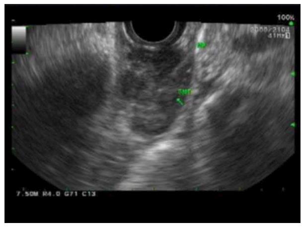
Figure 4 Endoscopic ultrasonography images of esophageal gastrointestinal stromal tumors.
A distal esophageal submucosal lesion measuring 2.6 cm × 1.3 cm in diameter was noted to be well circumscribed, heterogeneous with hypoechoic echotexture without disruption of wall architecture and with no perilesional lymph node.
- Citation: Lim KT, Tan KY. Current research and treatment for gastrointestinal stromal tumors. World J Gastroenterol 2017; 23(27): 4856-4866
- URL: https://www.wjgnet.com/1007-9327/full/v23/i27/4856.htm
- DOI: https://dx.doi.org/10.3748/wjg.v23.i27.4856


