Copyright
©The Author(s) 2017.
World J Gastroenterol. Jul 14, 2017; 23(26): 4767-4778
Published online Jul 14, 2017. doi: 10.3748/wjg.v23.i26.4767
Published online Jul 14, 2017. doi: 10.3748/wjg.v23.i26.4767
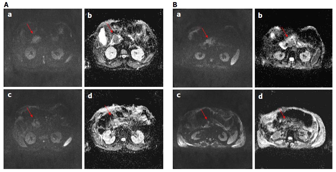
Figure 4 Diffusion weighted imaging assessment using morphological criteria for two patients (in A man 79 years old and in B man 74 years old).
In a (image at b value 800), in b (ADC map) is showed the lesion before the treatment and in c (image at b value 800) and d (ADC map) is showed the lesion after the treatment; there was a difference in diffusion maps before and after treatment.
- Citation: Granata V, Fusco R, Setola SV, Piccirillo M, Leongito M, Palaia R, Granata F, Lastoria S, Izzo F, Petrillo A. Early radiological assessment of locally advanced pancreatic cancer treated with electrochemotherapy. World J Gastroenterol 2017; 23(26): 4767-4778
- URL: https://www.wjgnet.com/1007-9327/full/v23/i26/4767.htm
- DOI: https://dx.doi.org/10.3748/wjg.v23.i26.4767


