Copyright
©The Author(s) 2017.
World J Gastroenterol. Jul 14, 2017; 23(26): 4767-4778
Published online Jul 14, 2017. doi: 10.3748/wjg.v23.i26.4767
Published online Jul 14, 2017. doi: 10.3748/wjg.v23.i26.4767
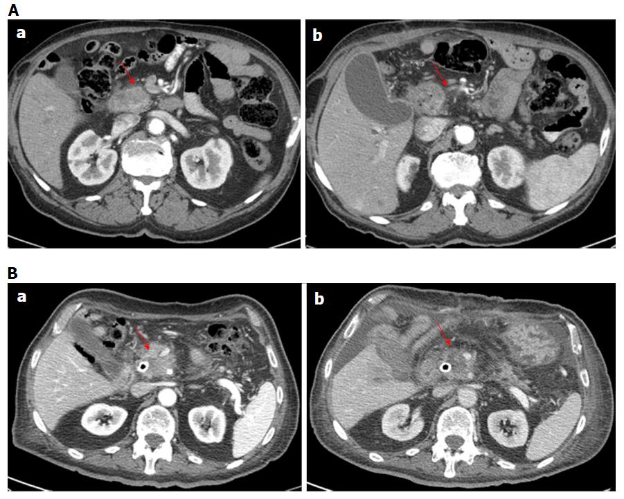
Figure 2 Computed tomography imaging assessment using morphological criteria for two patients (in A man 79 years old and in B man 74 years old).
In pancreatic phase on CT study (a) the lesion appears hypodense (arrow). After the treatment in pancreatic phase on CT study (b) the lesion appears similar than in (a) but there was a significant variation in CT density value. CT: Computed tomography.
- Citation: Granata V, Fusco R, Setola SV, Piccirillo M, Leongito M, Palaia R, Granata F, Lastoria S, Izzo F, Petrillo A. Early radiological assessment of locally advanced pancreatic cancer treated with electrochemotherapy. World J Gastroenterol 2017; 23(26): 4767-4778
- URL: https://www.wjgnet.com/1007-9327/full/v23/i26/4767.htm
- DOI: https://dx.doi.org/10.3748/wjg.v23.i26.4767


