Copyright
©The Author(s) 2017.
World J Gastroenterol. Jul 7, 2017; 23(25): 4615-4623
Published online Jul 7, 2017. doi: 10.3748/wjg.v23.i25.4615
Published online Jul 7, 2017. doi: 10.3748/wjg.v23.i25.4615
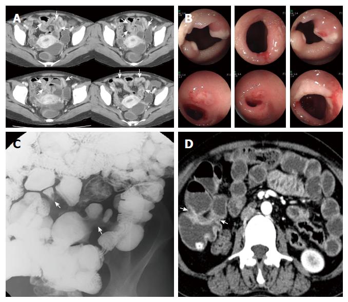
Figure 2 A 34-year-old woman with anemia.
A: Axial images of contrast-enhanced computed tomography (CT) enterography show segmental bowel wall thickening (arrows) with suspicious strictures along the distal ileum; B: On retrograde double-balloon endoscopic examination at the same time period there are multiple sharply demarcated ulcers at or near the strictures of the ileum; C: Small bowel series spot radiograph obtained two months later reveals segmental luminal narrowing (arrows) along the distal ileum, without overt ulceration; D: On axial images of contrast-enhanced CT enterography after two years, previous bowel wall thickening of distal ileum has improved and prominent strictures are noted at the corresponding ileal segment.
- Citation: Hwang J, Kim JS, Kim AY, Lim JS, Kim SH, Kim MJ, Kim MS, Song KD, Woo JY. Cryptogenic multifocal ulcerous stenosing enteritis: Radiologic features and clinical behavior. World J Gastroenterol 2017; 23(25): 4615-4623
- URL: https://www.wjgnet.com/1007-9327/full/v23/i25/4615.htm
- DOI: https://dx.doi.org/10.3748/wjg.v23.i25.4615


