Copyright
©The Author(s) 2017.
World J Gastroenterol. Jun 21, 2017; 23(23): 4252-4261
Published online Jun 21, 2017. doi: 10.3748/wjg.v23.i23.4252
Published online Jun 21, 2017. doi: 10.3748/wjg.v23.i23.4252
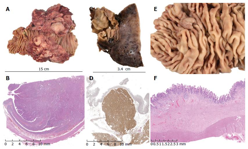
Figure 5 Pathological findings.
A and B: exophytic lesion in the duodenum shown to be a moderately differentiated duodenal adenocarcinoma on histology (haematoxylin and eosin stain); C and D: gastrointestinal stromal tumour confirmed on immunohistochemistry with CD117 and DOG1 staining; E and F: tubulovillous adenoma of the duodenum with low-grade dysplasia on histology (haematoxylin and eosin stain).
- Citation: Mitchell WK, Thomas PF, Zaitoun AM, Brooks AJ, Lobo DN. Pancreas preserving distal duodenectomy: A versatile operation for a range of infra-papillary pathologies. World J Gastroenterol 2017; 23(23): 4252-4261
- URL: https://www.wjgnet.com/1007-9327/full/v23/i23/4252.htm
- DOI: https://dx.doi.org/10.3748/wjg.v23.i23.4252


