Copyright
©The Author(s) 2017.
World J Gastroenterol. Jun 21, 2017; 23(23): 4252-4261
Published online Jun 21, 2017. doi: 10.3748/wjg.v23.i23.4252
Published online Jun 21, 2017. doi: 10.3748/wjg.v23.i23.4252
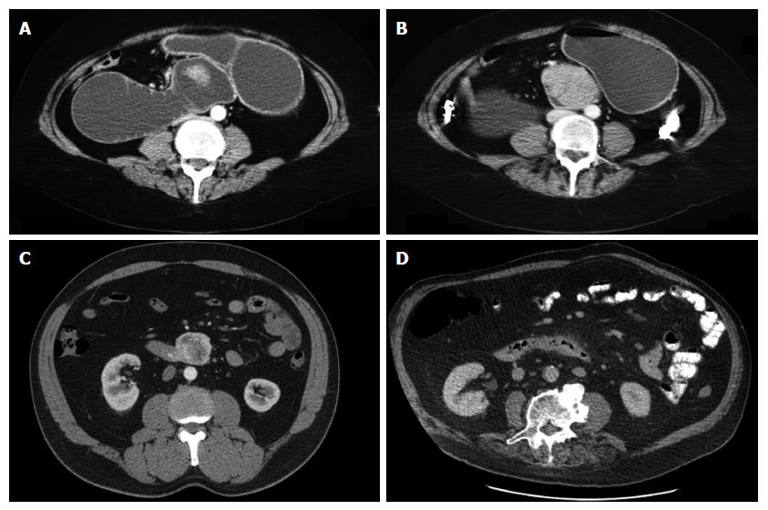
Figure 1 Representative axial computed tomography imaging of duodenal adenocarcinoma.
A and B: obstruction due to a large duodenal mass (same patient); C: exophytic mass without obstruction; D: subtle thickening of duodenum and periduodenal fat stranding reported as duodenitis, but in fact was a malignant tumour on post resection histology.
- Citation: Mitchell WK, Thomas PF, Zaitoun AM, Brooks AJ, Lobo DN. Pancreas preserving distal duodenectomy: A versatile operation for a range of infra-papillary pathologies. World J Gastroenterol 2017; 23(23): 4252-4261
- URL: https://www.wjgnet.com/1007-9327/full/v23/i23/4252.htm
- DOI: https://dx.doi.org/10.3748/wjg.v23.i23.4252


