Copyright
©The Author(s) 2017.
World J Gastroenterol. Jun 21, 2017; 23(23): 4211-4221
Published online Jun 21, 2017. doi: 10.3748/wjg.v23.i23.4211
Published online Jun 21, 2017. doi: 10.3748/wjg.v23.i23.4211
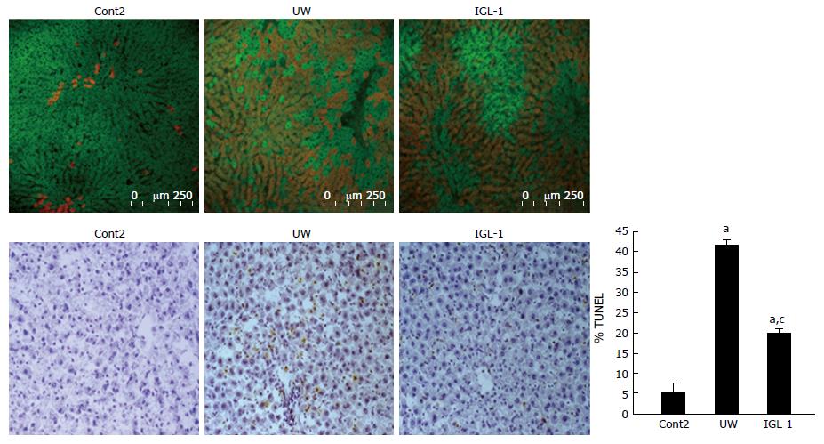
Figure 6 Confocal microscopy findings and Tunel assay after reperfusion.
Confocal microscopy images showing green fluorescence of rhodamine 123 cell viability marker and Evans blue dye (in red) used as a viability assay on the basis of its penetration into non-viable cells. Representative light photomicrographs of TUNEL-stained sections. Steatotic livers submitted to cold storage preservation with UW showed numerous positive cells (both hepatocytes and sinusoidal lining cells) compared with control non-preserved livers. The positivity decreased when the livers were submitted to cold storage preservation with IGL-1 solution. aP < 0.05 vs Cont2, cP < 0.05 vs UW. Scale bar 50 μm. Cont2: Liver flushed and perfused ex vivo without cold preservation; UW: Liver preserved in UW solution; IGL-1: Liver preserved in IGL-1 solution; UW: University of Wisconsin; IGL-1: Institut Georges Lopez-1.
- Citation: Zaouali MA, Panisello-Roselló A, Lopez A, Castro Benítez C, Folch-Puy E, García-Gil A, Carbonell T, Adam R, Roselló-Catafau J. Relevance of proteolysis and proteasome activation in fatty liver graft preservation: An Institut Georges Lopez-1 vs University of Wisconsin appraisal. World J Gastroenterol 2017; 23(23): 4211-4221
- URL: https://www.wjgnet.com/1007-9327/full/v23/i23/4211.htm
- DOI: https://dx.doi.org/10.3748/wjg.v23.i23.4211


