Copyright
©The Author(s) 2017.
World J Gastroenterol. Jun 21, 2017; 23(23): 4181-4190
Published online Jun 21, 2017. doi: 10.3748/wjg.v23.i23.4181
Published online Jun 21, 2017. doi: 10.3748/wjg.v23.i23.4181
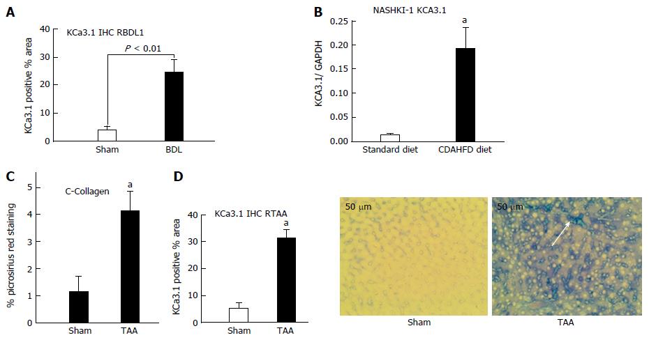
Figure 1 Upregulation of KCa3.
1 expression in diseased livers. A: In rats subjected to biliary occlusion, hepatic KCa3.1 expression, probed with anti-KCa3.1 (SK4, IKCa1) antibody, was several-fold that in of sham operated rats. B: Densitometric analysis was made of Western blots for KCa3.1 expression in liver lysates from mice fed a standard diet or CDAHFD for 8 wk. Compared to its expression in livers from animals on a standard diet, KCa3.1 expression is increased several-fold in livers from the CDAHFD cohort (aP < 0.05 vs standard diet). C: Left: Livers from rats administered TAA for 8 wk exhibited increased fibrosis evidenced by increased picrosirius red staining (aP < 0.05 vs sham). D: Increased KCa3.1 expression significantly (aP < 0.05 vs Sham) (middle). (20 ×), intense blue membranous staining (white arrow and right bottom) was noted in these fibrotic livers.
- Citation: Paka L, Smith DE, Jung D, McCormack S, Zhou P, Duan B, Li JS, Shi J, Hao YJ, Jiang K, Yamin M, Goldberg ID, Narayan P. Anti-steatotic and anti-fibrotic effects of the KCa3.1 channel inhibitor, Senicapoc, in non-alcoholic liver disease. World J Gastroenterol 2017; 23(23): 4181-4190
- URL: https://www.wjgnet.com/1007-9327/full/v23/i23/4181.htm
- DOI: https://dx.doi.org/10.3748/wjg.v23.i23.4181


