Copyright
©The Author(s) 2017.
World J Gastroenterol. Jun 14, 2017; 23(22): 4127-4131
Published online Jun 14, 2017. doi: 10.3748/wjg.v23.i22.4127
Published online Jun 14, 2017. doi: 10.3748/wjg.v23.i22.4127
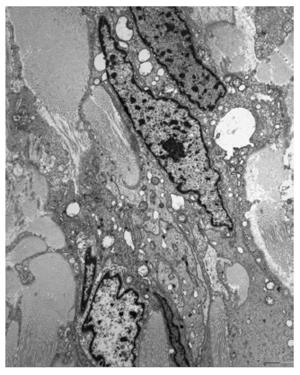
Figure 4 Electron microscopic findings of the submucosal tumor.
Elongated neoplastic cells show with numerous complex interdigitating cytoplasmic processes. The cytoplasmic membrane was completely covered with external lamina. The nuclei reveal irregular margins and a heterochromatic chromatin pattern, and the perikaryal cytoplasm contained several mitochondria, rough endoplasmic reticulum, ribosomes, and many lysosomes.
- Citation: Choi KW, Joo M, Kim HS, Lee WY. Synchronous triple occurrence of MALT lymphoma, schwannoma, and adenocarcinoma of the stomach. World J Gastroenterol 2017; 23(22): 4127-4131
- URL: https://www.wjgnet.com/1007-9327/full/v23/i22/4127.htm
- DOI: https://dx.doi.org/10.3748/wjg.v23.i22.4127


